By Natalie Nielsen
Fun rating: 5/5

Difficulty rating: 4/5

What is the general purpose? To visually label and detect proteins on and in cells.

Why do we use it? Medical diagnostics or scientific research. For example, it can aid in the diagnosis of breast cancer.
How does it work? Have you ever wondered how scientists identify specific cell types? At the macroscopic level, where objects are visible with the naked eye, it is easy for us to distinguish between different tissues- like skin and blood. Within tissues, however, there are many different cell types with specialized functions that are only visible microscopically. For example, in a tumor there are cancer cells, immune cells, blood vessels, lymphatic vessels, fibroblasts, etc. Scientists and doctors use different techniques to see and study these cells.

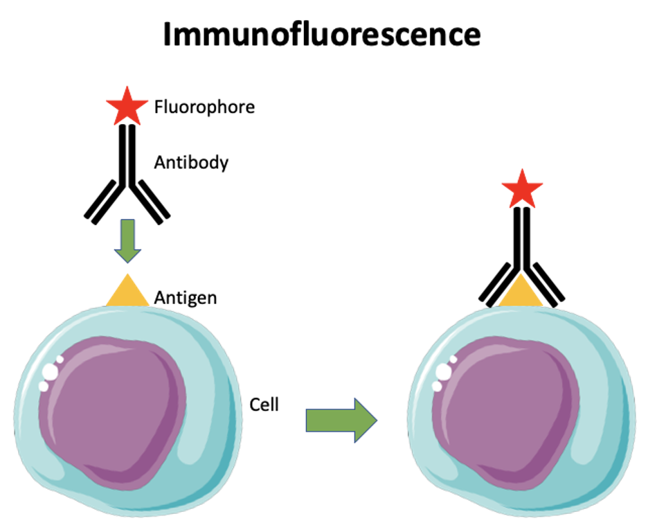
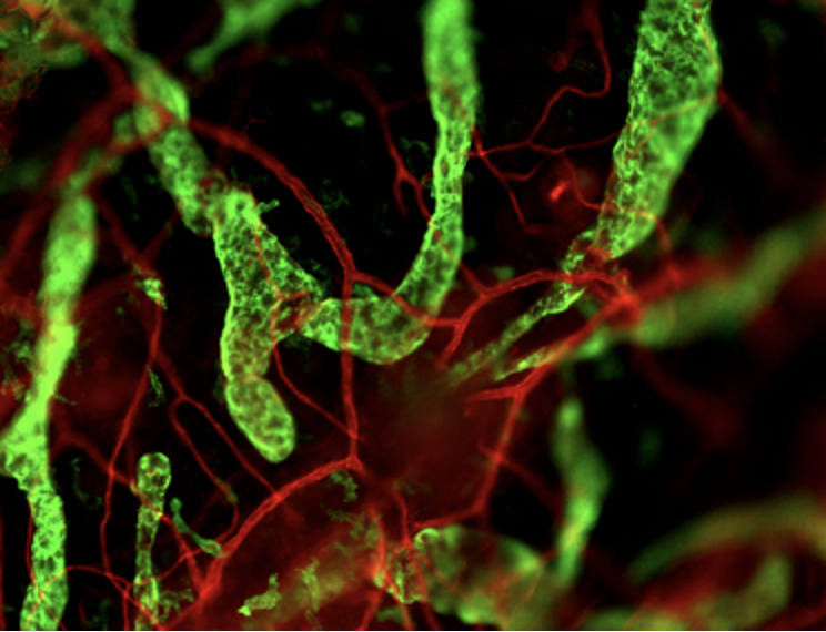
One of the most common methods used to visually identify cells within a tissue is called immunofluorescence. Despite its long name, it is a simple technique that generates eye-catching images, like the examples provided in the article. The first step involves acquiring a tissue of interest such as ear, heart or a tumor biopsy. This tissue is then “fixed” with formaldehyde (yes, the smelly, toxic liquid you may be familiar with from biology class) to cross-link proteins and prevent the tissue from decaying. The fixed tissue is then sliced into very thin sections, and placed on a glass slide. Next, slides are immersed in a solution containing an antibody. Antibodies are big, Y-shaped proteins that specifically recognize one “antigen” (the single protein you are looking for) out of the hundreds of thousands that exist in animal cells. At the top of the antibody is a fluorophore (a protein that can emit light in a range of colors) which can be red, green, or blue and provides the color in the images you see here. The bottom of the antibody will bind to the antigen, the single protein of interest. This technique provides colorful images that allow visualization of specific cell types, which is useful in evaluating disease and for other research.


