by Deepika Jayaprakash
Technique Name: Metaphase spreads
Fun Rating: 4/5

Difficulty Rating: 4/5
This technique is invaluable for clinical diagnosis and research. However, isolating cells of interest from clinical samples and growing them in the lab is difficult, and, as if this wasn’t complicated enough, obtaining good quality metaphase spreads is technically challenging.
What is the purpose and why do we use it?
Metaphase spreads are used in cytogenetics (the study of chromosomes) to study the relationship between chromosomal abnormalities and human genetic diseases. Indications for this laboratory test include, but are not limited to
- Genetic evaluation of an unborn child (prenatal) to rule out chromosomal defects in early pregnancy
- Postnatal diagnosis of developmental delay and congenital anomalies in infants and children
- Chromosomal abnormalities in cancers
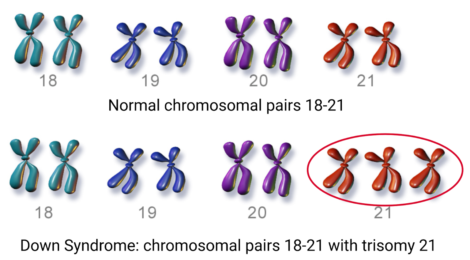
A normal human cell contains 23 pairs of chromosomes, including 22 pairs of autosomes and one pair of sex chromosomes (XX or XY). Any deviation from 23 pairs and the well-established structure of human chromosomes is associated with various diseases. For example, a discrepancy in chromosome numbers is called aneuploidy which involves either
- Missing chromosomes or
- One or more extra chromosomes
The most common aneuploidies are Down syndrome with trisomy 21 (extra copy of chromosome 21- Figure 1) and Turner syndrome with monosomy X (missing or partially missing one of the X chromosomes).
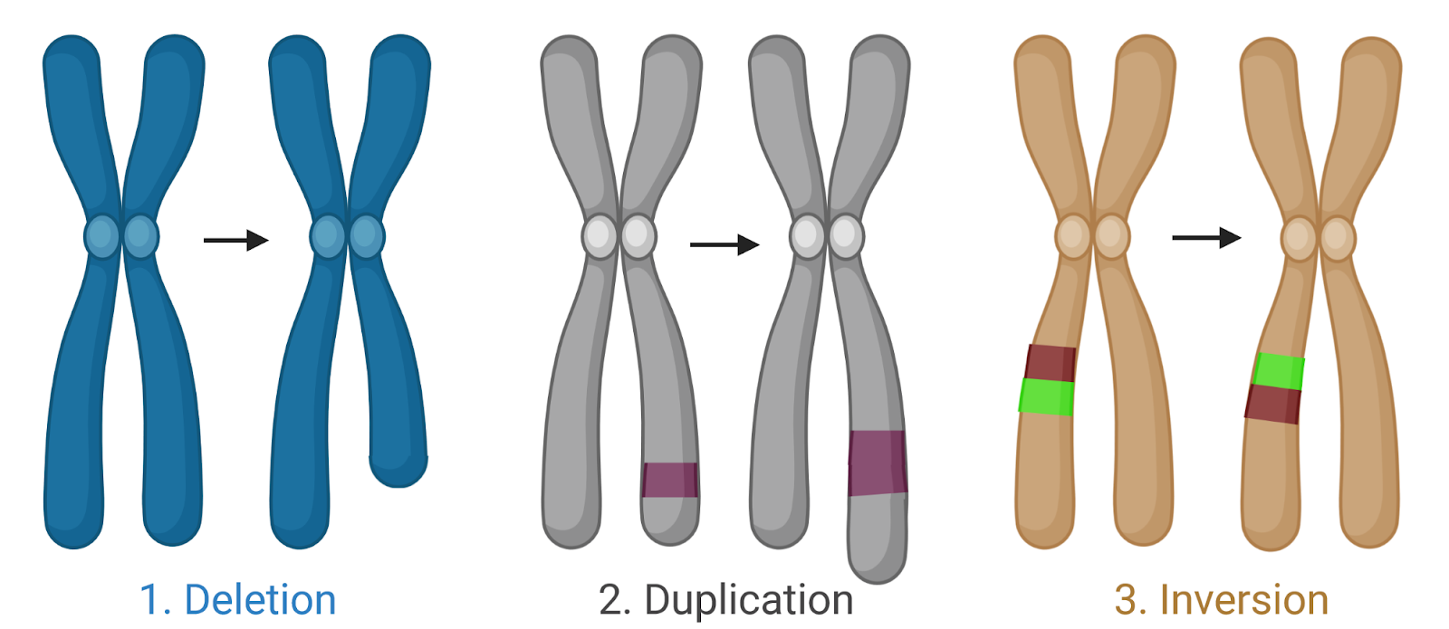
Chromosomal structural abnormalities include (Figure 2)
- Deletion: Part of a chromosome is missing
- Duplication: Part of a chromosome is repeated
- Inversion: Part of the chromosome is inverted within the chromosome
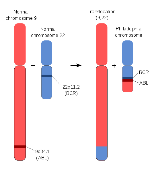
- Translocation: Material between two different chromosomes is exchanged (Figure 3)
- Insertion: Addition of material from another chromosome.
Metaphase spread technique helps investigate such deviations from normal chromosomal numbers and structure.
How does the metaphase spread technique work?
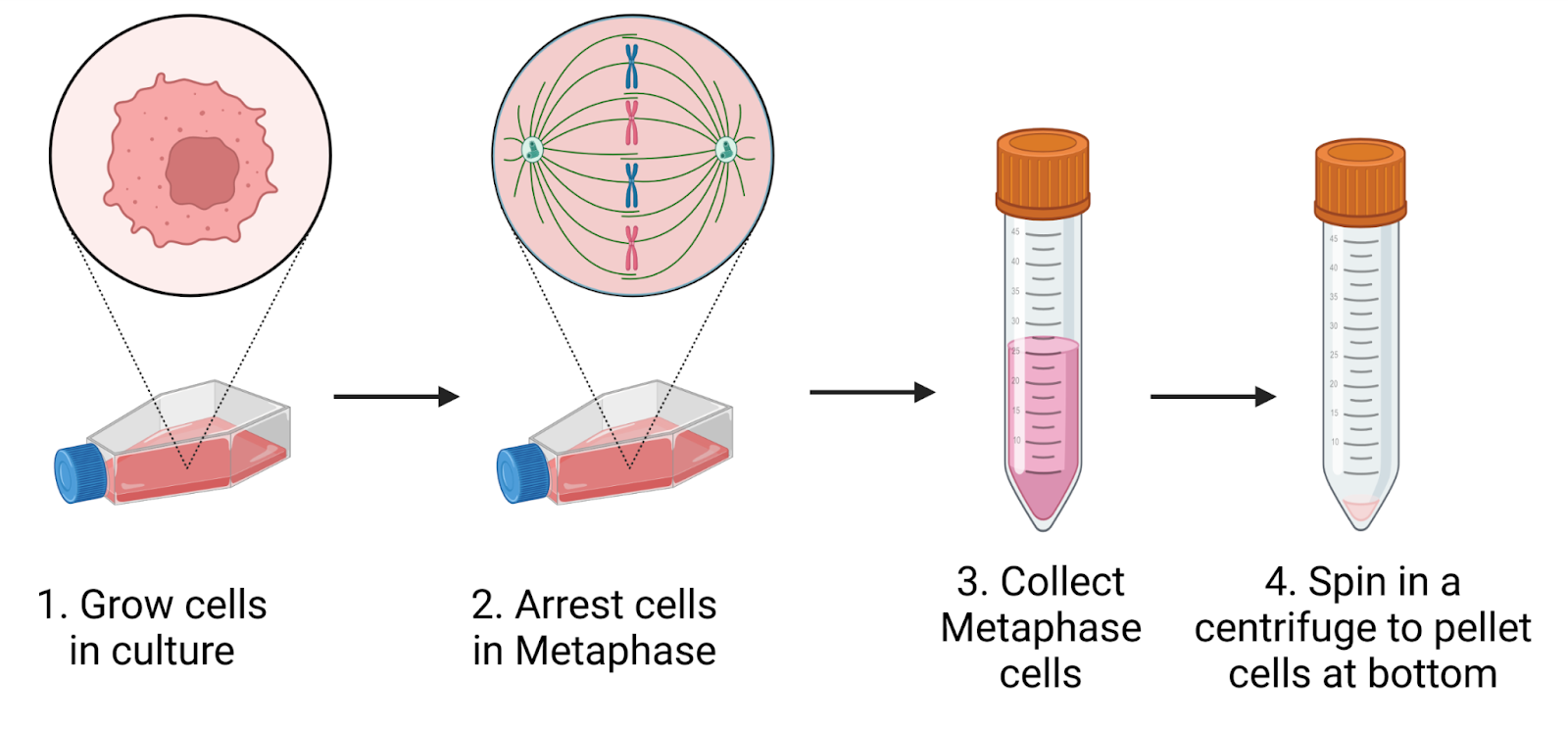
DNA in human cells exists in complex with various proteins, forming long strands of chromatin within the nucleus. During cell division/mitosis, particularly during metaphase, the long-coiled chromatin condenses into chromosomes that can be visualized under the microscope. Therefore, in the metaphase spread technique, cells isolated from clinical samples are cultured and treated with an agent (eg: colcemid, Nocodozole) that arrests the cells in metaphase (Figure 4).
Once the cells arrested in metaphase are harvested, they are treated with a hypotonic solution (solution with lower osmotic pressure and lower solute concentration than the cells) so that water enters the cell and causes them to swell (Figure 5). The swollen cells are fragile (therefore should be handled with care) and hence can easily rupture when dropping them on the slide at a later stage (step 8). The sample is then fixed–a preservation technique used to prevent degradation by internal and external environmental factors–by adding a freshly prepared mixture of 3:1 Methanol to acetic acid fixative. Once cells are fixed, they can be stored for several months before dropping them onto slides for observation.

When dropping the samples onto slides, the samples should be resuspended in fresh fixative. The slide should then be immediately flooded with the fixative solution (Figure 6). As the fixative flows and dries, it stretches the ruptured metaphase chromosomes on the slide. Temperature and humidity affect the time required for the fixative to dry, thus affecting the quality of the spread. For best results, it is important to optimize for the environmental conditions every day this experiment is performed. Once the slides dry, they can either be stained by DAPI/Giemsa stain to visualize the chromosomes (Figure 7) or processed further to evaluate structural details more closely.


The analysis of metaphase spreads can be done either manually or by using various software tools like ChromaWizard, IdeoKar or MetaSel. When I did my first metaphase spread analysis, I sat in the dark room for 6 days to quantify all my slides under the microscope. Although I still analyze manually, I have streamlined the process to be completed within 3 days!
To conclude, metaphase spread analysis serves to establish diagnosis of genetic diseases, and identify chromosomal defects. In the case of cancer studies, chromosome analysis can provide insight into alterations in specific biological pathways. Defining such alterations helps to identify specific drugs/inhibitors that can destroy cancer cells and serve in cancer treatment.
