by Karly Forker
Fun Rating: 4/5
Difficulty Rating: 2/5
What is the general purpose?
Size exclusion chromatography is a technique used to separate molecules in a solution based on their size or molecular weight.
Why do we use it?
Scientists may use size exclusion chromatography to separate molecules from one another for purification later or to analyze the molecular weight distribution of a sample.
How does it work?
- Preparing the column
The size exclusion chromatography column looks like a long cylindrical tube and it is filled with tiny porous beads made of materials like agarose or silica. Though the size of the bead differs based on the column type, an individual column’s beads will all be the same size. Before applying your sample, a buffer is typically used to wash the column to make the sample feel more at home when it is applied.
- Sample application
A small volume of your sample is applied to the column, oftentimes by injecting it onto a loop which acts like a waterslide delivering your sample to the top of the column.
- Sample flow and separation
More buffer is applied to help push the sample down the column. This buffer is called the mobile phase. As the sample flows down the column, different-sized molecules interact with the beads in different ways. Large molecules are unable to fit inside the beads’ pores, and so move quicker down the column. Conversely, smaller molecules can enter the pores of the beads before continuing their flow down the column, causing them to move slower.
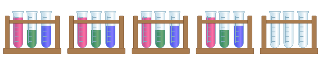
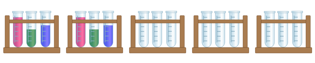
- Fraction collection
As the liquid exits the column, it is collected in small segments called fractions in order to separate the bigger molecules (earlier fractions) from the smaller ones (later fractions).
- Analyzing results
In order to determine what molecules are in what fractions, scientists may run a Coomassie gel (a gel used to visualize proteins based on their molecular weight) or gel electrophoresis in order to visualize the size of the molecules in each fraction to get an idea of the distribution of the sample.
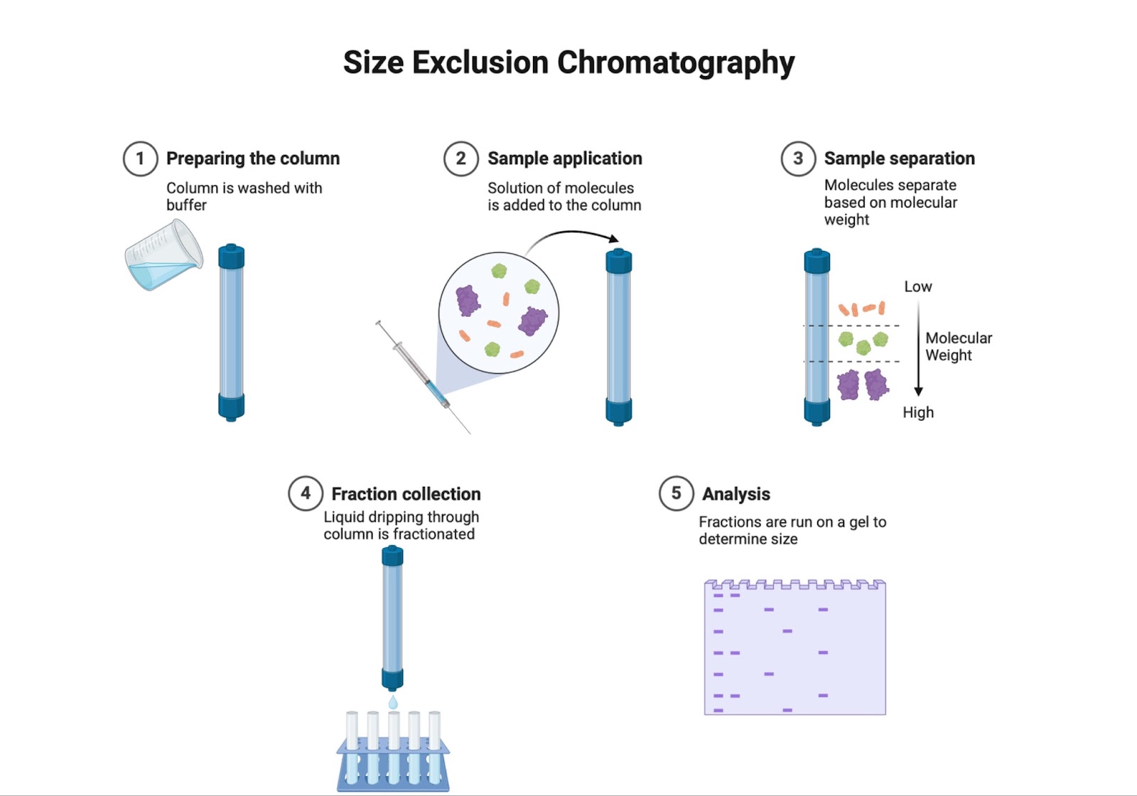
Figure 1: The process of size exclusion chromatography. Image made by author using BioRender.
