by Jessica Zhang
Fun Rating: 5/5
Difficulty Rating: 3/5
What is the general purpose? Neuronal tracing allows neuroscientists to identify the start and end points of a group of neurons. Studying the connectivity of neurons contributes to our understanding of brain anatomy and can help us to infer their functions.
Why do we use it? The brain is a highly complex structure. Think about the complex tasks that it manages for you every day, including reading this post! To carry out these complex functions, the brain is organized into specialized regions, and coordination within and between regions is crucial. When reading and learning from NC DNA Day Blog posts, for example, you will need to engage your visual cortex located at the back of your brain, your prefrontal cortex located at the front of your brain, and many other areas in between to receive the visual input, process the semantic meaning of sentences, link new pieces of knowledge to your memory… A major class of neurons, known as excitatory neurons, are tasked to manage communications within and between regions in the central nervous system through transporting electrical signals generated from one place to another. Anatomically, they receive electrical signals through structures known as dendrites in the soma, and they send these signals along the axon, which then reaches the axon terminals on the other end of the neuron and passes along the signal to other neurons that are connected (Figure 1).
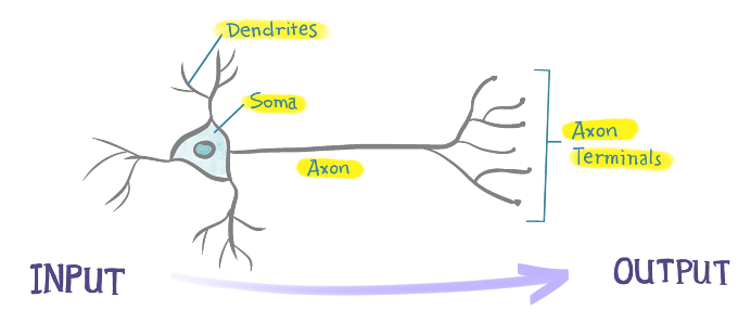
Figure 1: Structures in an excitatory neuron. The arrow represents the direction of electrical signal transduction.
It is important for neuroscientists to know the start and end point of a neuron, since we can infer their functional relevance through their anatomy. Let’s say we have found a group of neurons that send signals from the visual cortex to the prefrontal cortex. Maybe you can then formulate a hypothesis and test for it: are these neurons involved in reading comprehension?
How does it work?
Types of tracing
There are broadly two types of tracing:
1. Anterograde tracing (forward tracing)
The tracer is taken up by the soma and travels along the axon to reach the axon terminals. Forward tracing identifies regions where neurons SEND information to.
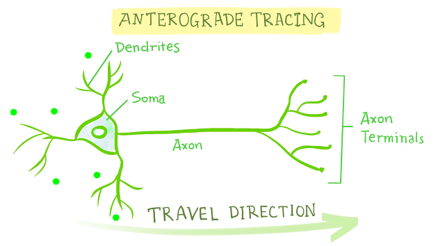
Figure 2: Anterograde tracing. The arrow represents the direction of the tracers traveling along the axon.
2. Retrograde tracing (backward tracing)
The tracer is taken up by the axon terminals and travels along the axon to reach the soma. Backward tracing identifies regions where neurons RECEIVE information from.
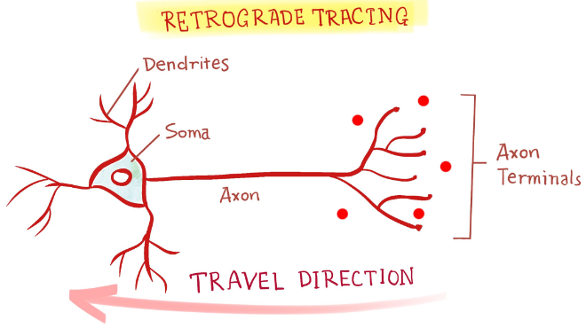
Figure 3: Retrograde tracing. The arrow represents the direction of the tracers traveling along the axon.
Tracers
Traditional tracers include proteins that are transported in a certain direction along the axon, like PHA-L (a plant protein) for anterograde tracing and CTB (a bacterial protein) for retrograde tracing. They are engineered to be coupled with fluorescent proteins, so the entire neuron lights up. Recently, it is becoming increasingly common for neuroscientists to use AAV (adeno-associated virus) to perform neuronal tracing. AAV generates long-lasting signals (weeks to months), has minimal cytotoxicity, and has various serotypes for both anterograde and retrograde tracing. Most importantly, AAV-based tracing allows conditional expression, which adds another layer of control to the neuronal population labeled. Let’s say you are interested in the connectivity of neurons expressing a certain gene of interest. AAV-based tracing allows the possibility of only expressing signals if this gene is present. Therefore, when you look at the brain after injection, only those with the gene of interest light up.
Procedure
The experimental process is very straightforward. The tracers are injected into the region of interest in the brain through stereotaxic injection, which enables injection at a precise coordinate in the brain. After waiting for some time to allow for expression, the brains are harvested to be sectioned and imaged under the microscope. Then enjoy the fun of identifying interesting brain areas!
