by Ana Cunningham
Fun Rating: 4/5

Difficulty Rating: 2/5

What is the general purpose?
The lactate dehydrogenase (LDH) assay is a cytotoxicity (cell death) /cell viability assay that measures the proportion of dead cells in culture. Cell death is quantified by measuring the light emitted as a byproduct of secreted enzymes (LDH) after the cell dies.
Why do we use it? When we do experiments in cell culture, it is vital to know which cells are alive and which are dead, whether your goal is to see if a treatment induces cytotoxicity or to exclude dead cells from your experiment. The LDH assay is one of the most popular cytotoxicity assays because it is simple to perform and perfect for high-throughput screening, meaning you can measure the cytotoxicity of many cell populations at once, such as by screening an entire 96-well plate of cells (Figure 1). Because the assay is performed on the media rather than the cells themselves, you can preserve your model for other measurements, such as RNA or protein extraction.
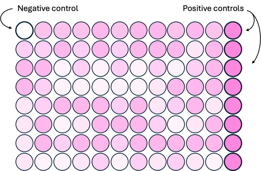
Figure 1. Example of high-throughput screening for LDH cytotoxicity assay in a standard 96-well plate. Darker shades of pink indicate higher levels of cytotoxicity. The rightmost column represents positive controls in which all samples are lysed, and the top-left well represents a negative control. Created by author using BioRender.
How does it work?
When cells die, their membrane (outer area) becomes permeable, meaning that all the enzymes and other contents inside the cell can flow out into the surrounding media. One of these enzymes, lactate dehydrogenase (LDH), triggers a reaction that produces light as a byproduct, and the LDH assay takes advantage of this by measuring light produced as a proxy for cell death, also known as cytotoxicity. See more about the chemical reactions involved in the LDH assay here. In short, scientists can add a reagent called INT to the cell media so that when LDH enters the media, it interacts with INT and indirectly creates a fluorescent product called formazan (Figure 2). Here’s how:
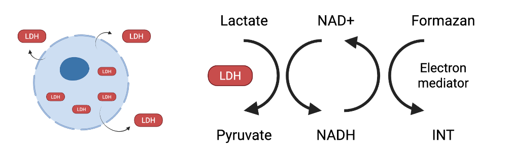
Figure 2. Schematic of LDH cytotoxicity assay mechanisms of action. Damaged cells secrete the enzyme LDH, which converts lactate to pyruvate and produces NADH as a byproduct. NADH reduces iodonitrotetrazolium chloride (INT) to formazan, which emits red light that can be quantified by a colorimeter as a proxy for cell death. (Created by author in Biorender and modified based on source: Creative Bioarray).
- INT and its converting enzyme (diaphorase) are added to small amounts of media from the cell culture of interest. These components, in addition to chemicals already present in the media, are all you need to generate the light detected by the colorimeter.
- Note! Whenever you perform an experiment, you should have positive and negative controls. In this case, your positive control contains media from a group of 100% dead cells. This represents the maximum absorbance, or the maximum amount of LDH that can be secreted. There should also be a negative control containing only water or unused cell media to measure background absorbance.
- The mixture is incubated for 15 minutes at room temperature away from light. This ensures that photobleaching does not occur, which can limit the accuracy of the assay.
- After 15 minutes, the wells will be visibly pink. The plate is inserted into a colorimeter (a machine that measures specific wavelengths, or colors, of light), which measures the absorbance of the light emitted by formazan.
Other types of LDH assays
LDH assays can also be performed by triggering other chemical reactions that emit light or other detectable byproducts. For example, other applications of the LDH assay use an enzyme called luciferase, which converts luciferin into a product that emits another type of light. Certain variations of the LDH assay may be better suited for different applications. Besides LDH assays, there are many other ways to measure cytotoxicity that do not use LDH, including measuring the release of other enzymes (e.g., G6PD assay), gene expression assays (e.g., by PCR), dye uptake assays (e.g., Trypan blue), and more! With a diverse array of cytotoxicity assays at your disposal, you can be confident that the cells you measure are viable, thereby amplifying the strength of your experiment.
