by Yasemin Cole
Technique Name: Immunohistochemistry, abbreviated IHC
Fun Rating: 3/5
Difficulty rating: 4/5

What is the general purpose?
The goal of immunohistochemistry is to evaluate the location and quantity of a particular protein in a sample of tissue. It can be used in research and in clinical settings to diagnose diseases.
Why do we use it?
When performing research, we are dependent on model systems to understand the molecular world. Some researchers focus on animal models such as the common mouse (Latin name Mus musculus), whereas others use cells grown in the lab. Other posts have described techniques such as co-immunoprecipitation or western blotting which allows us to study proteins in all cells in the sample. Immunohistochemistry is different because it allows you to visualize proteins inside individual cells.

How does it work?
- Sample Collection:
To perform immunohistochemistry (IHC), a scientist first needs to collect either tissue or cells. The samples are treated with chemicals to prevent degradation and cell death. Typically we use paraformaldehyde, which is a chemical that binds proteins and DNA to each other to stabilize the molecules and prevent them from degrading. Because tissue is larger and more fragile, it is typically dehydrated to remove excess water and then embedded in a wax called paraffin (that helps to maintain the structure of the tissue) before it is sectioned into fine layers. A machine, called a microtome, slices the tissue into small 5 micrometer sections (that is approximately ½ of the diameter of a mammalian cell).
- Antibody incubation
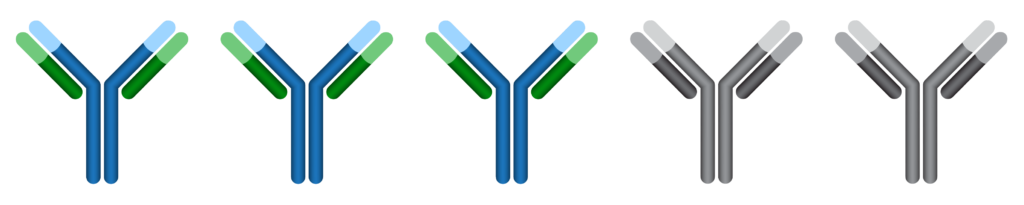
The samples are next incubated in an antibody that is designed to specifically bind to the protein of interest. You may have heard the concept of a molecule binding to a protein like a lock and a key. Similarly, the antibody binds to a specific region of the protein as seen in the “Immunohistochemistry Basics” figure.
- Signal Amplification and Detection
Typically secondary antibodies are used to amplify the signal. Similar to the primary (first) antibody, a secondary antibody binds to a specific region of the first antibody. This second antibody is special because an enzyme is attached to this antibody that initiates a color-changing chemical reaction. Using this color-based detection system, the enzyme converts 3,3′-diaminobenzidine into a brown precipitate that can be seen with a microscope. In effect, in the locations where the primary antibody is not present, no color change will be produced. A visible colored precipitate will be produced in areas where the primary antibody binds to the tissue.

How is it used?
Immunohistochemistry is a staple of modern biological science and is used in medicine to diagnose diseases. For example, IHC helps us determine the cancer subtype of a newly diagnosed cancer patient. When an individual is diagnosed with breast cancer, cells in their biopsy sample are stained to determine whether the cancer cells express proteins called estrogen receptor, progesterone receptor, or human epidermal growth factor type 2. Depending on the results, patients will receive personalized therapies targeting the above proteins.
