by Megan (Frederick) Amason
Fun Rating: 5/5

Difficulty Rating: 3/5
Technique name: TUNEL Staining (TUNEL = terminal deoxynucleotidyl transferase dUTP nick end labeling)
What is the general purpose? TUNEL staining labels partially degraded DNA that is generated within dying cells. Therefore, TUNEL staining enables detection and quantification (counting) of dying cells.
Why do we use it? In many instances, scientists want to identify cells that are undergoing apoptosis, or programmed cell death. TUNEL staining is one method used to identify cells that are dying.
How does it work? Did you know that the majority of the cells in your body are replaced every 7 years? While new cells arise through cell division, or mitosis, old cells are eliminated through cell death, or apoptosis. Apoptosis is essential during many processes: development, tissue and organ maintenance, and the elimination of potentially dangerous cells. Cells are considered dangerous when they accumulate changes (also known as mutations) in their genetic code, or DNA, because these changes can have negative consequences. In fact, most cancers are associated with mutations in DNA that cause cells to divide uncontrollably and resist apoptosis. Therefore, monitoring the ability of cells to undergo apoptosis, and sometimes directly causing cells to undergo apoptosis, is an important area of research.
How can researchers tell when a cell is dying by apoptosis? During apoptosis, several different events occur. First, enzymes known as caspases are activated and then cut or cleave many target proteins. This initiates a ripple effect of events that lead to DNA fragmentation, cell shrinkage, and membrane disassembly into small pieces known as apoptotic bodies (Figure 1). The ultimate goal of these events is to dispose of cells in a way that does not trigger the immune system to cause inflammation. By disposing of cells in a non-inflammatory manner, apoptosis maintains homeostasis in tissues despite the presence of cell death.

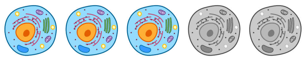
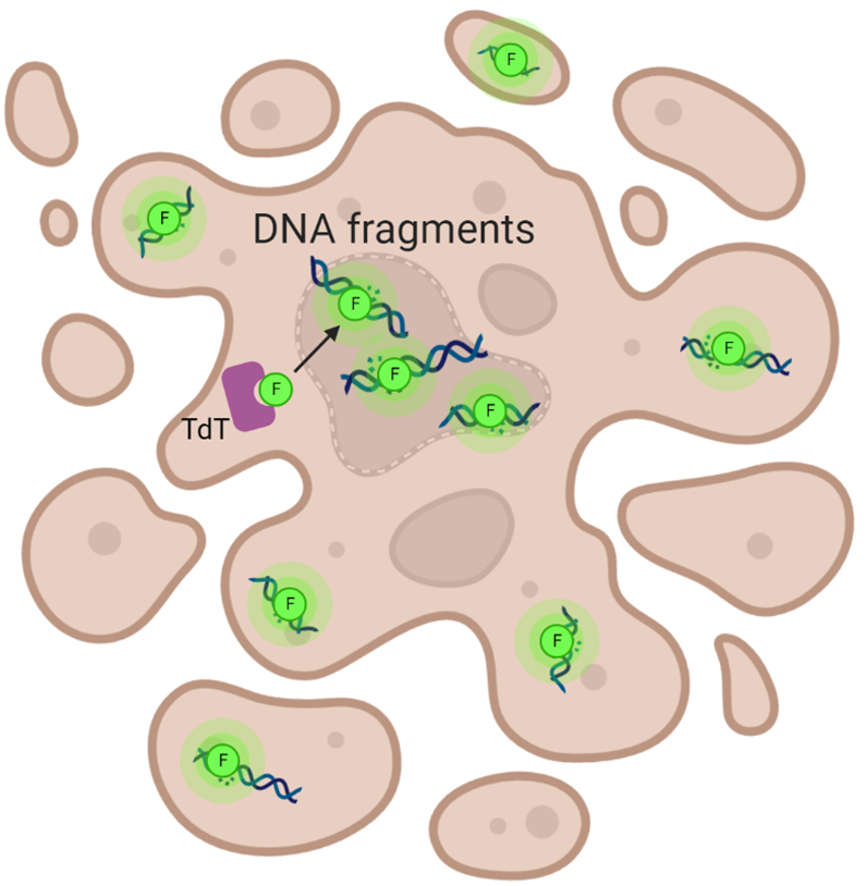
Many apoptotic events mentioned above can be detected by various methods, including TUNEL staining, which directly measures DNA fragmentation. TUNEL staining uses the enzyme TdT to label broken DNA within apoptotic cells (Figure 2). The broken DNA is usually labeled with fluorescent proteins that emit light when hit with a special laser. Another noteworthy aspect of apoptosis is breakdown of the nuclear envelope, an area that normally houses and protects DNA in healthy cells. Breakdown of the nuclear envelope allows fragmented DNA to leak into the cytosol of the cell. For this reason, fragmented DNA can be detected in apoptotic bodies. Once treated with TdT and the fluorescent label, apoptotic cells can be easily distinguished from healthy cells using a microscope or other fluorescent detection methods (Figure 3).

When do scientists use the TUNEL staining method? Suppose a scientist wants to find a new therapy that causes apoptosis of cancer cells. The scientist could treat cancer cells with different therapeutic drugs, then use TUNEL staining to measure apoptosis and determine the relative effectiveness of each drug in killing the cancer cells. Based on Figure 4, which drug is most effective at killing cancer cells? Which drug is least effective?
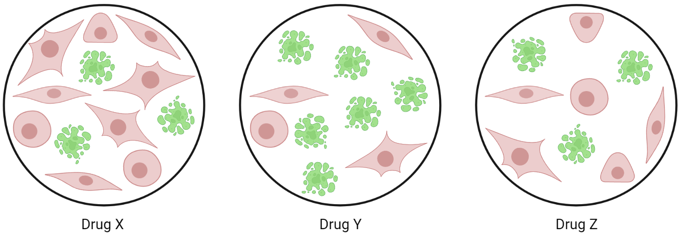
After a quick glance at the cells, the scientist is able to identify Drug Y as the most effective drug; the majority of cells treated with Drug Y are undergoing apoptosis. However, the difference between Drug X and Drug Z is less apparent. For the cells treated with Drug X, there are 3 cells undergoing apoptosis and 9 cells that look healthy, meaning that 25% of the cells are dying. For the cells treated with Drug Z, 3 cells are undergoing apoptosis and only 6 cells look healthy, meaning that about 33% of the cells are dying. By doing these calculations, the scientist is able to conclude that Drug X is the least effective at killing the cancer cells. This is one way a scientist can screen many drugs in many different types of cancer to reveal new therapies with the potential to treat cancer patients!
