by Margaret Dedloff
Technique name: Cytotoxicity assay or lactate dehydrogenase (LDH) release assay
Fun Rating: 4/5

Difficulty Rating: 2/5

What is the general purpose? Cytotoxicity assays, specifically lactate dehydrogenase (LDH) release assays, are used to measure cell death in culture. The main question that LDH release assays are used to answer is “what percentage of cells in this culture are dying due to the treatment they are receiving?”
Why do we use it? When giving cells with new drugs or infecting cells with bacteria or viruses, it is important to know how many cells are dying as a result of the drugyou added. LDH release assays can provide a measure of how effectvea drug is and how pathogenic bacteria or viruses are. Pathogenic bacteria/viruses are those that can cause disease. Bacteria/viruses that cause more severe disease (or in the case of a LDH release assay, more cell death) are more pathogenic. LDH release assays have very clear read-outs, making them an easy way to determine how many cells are dying in a culture. You can know if your drug has a toxic effect on cells in a matter of hours!
How does it work?
The LDH release assay is easy to use and produces a colorimetric change, meaning that the color of the media changes based on how much cell death there is (figure 1).

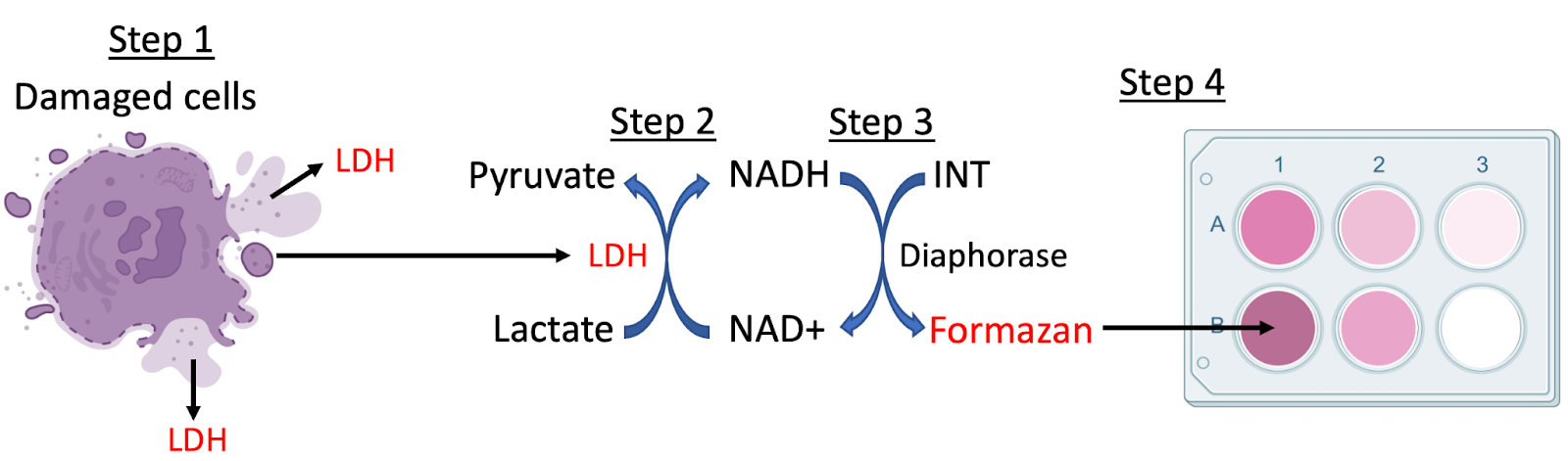
LDH is an enzyme present in nearly all mammalian cells. When tissues are damaged, cells release LDH into the media (Figure 2, Step 1). The LDH release assay has lactate in the media, which is converted to pyruvate by LDH (Step 2). This reaction produces NADH (Step 2). NADH is a cofactor, a molecule that helps enzymes, like diaphorase, to complete their functions. Using NADH, diaphorase converts INT into formazan (Step 3), which produces the colorimetric change in the media (Step 4). INT is a salt that can be reduced to formazan, a red pigment.
Stage 1: Mammalian cells are grown in culture to a known number of cells. It is important to make sure that each sample you have contains the same number of cells so that at the end of the assay (step 5), you can calculate the percentage of cell death.
Stage 2: Cells are treated with drugs or infected with bacteria or viruses. Cells are left to incubate with their treatment for a predetermined amount of time. One group of cells are treated with Triton X-100, a chemical that causes cell death, to make sure that all cells in that group die. This is a positive control. Another group of cells should not be treated with anything. This is a negative control. These two groups will allow you to calculate how much cell death is experienced by your treated groups (step 5).
Stage 3: A small portion of liquid media is collected and incubated with lactate. If LDH is present from cells that have died, it converts lactate to pyruvate. This produces NADH, which diaphorase uses to convert INT to formazan, producing a red color change.
Stage 4: The amount of color change can be measured by measuring the optical density of the sample using a plate reader.
Stage 5: Calculate amount of cell death for sample using the following equation:

