by Leslie Kent
Technique Name: Crystal Violet Biofilm Assay
Fun Rating: 5/5
Difficulty Rating: 2/5
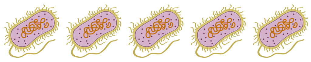
What is the general purpose? Microbes are living organisms so small that we need to use a microscope to see them, such as bacteria and fungi. However, these tiny organisms can work together to form bigger group structures called biofilms. Biofilms are community structures that are attached to a surface surrounded by a protective layer made up primarily of sugars. Biofilms can help microbes survive as a community in environments that could be dangerous for them if they were growing as single cells, such as when infecting a host. Microbiologists (scientists who study microbes), can use a technique called the crystal violet biofilm assay to measure biofilms.
Why do we use it? Many types of microbes make biofilms. Biofilms protect microbes from environmental stress, antimicrobial treatments like antibiotics, and immune system responses. Understanding how different microbes form biofilms is important so we can learn how to kill microbes that cause problems such as decreased crop yield in agriculture, dental plaque on our teeth that can result in disease, and persistent infections that are more difficult to treat in humans and other animals.
How does it work? After scientists pick out what type of microbes they want to study, they add microbes to the wells of a microtiter plate, a specialized plate with many small wells. You can see these wells in the picture below.
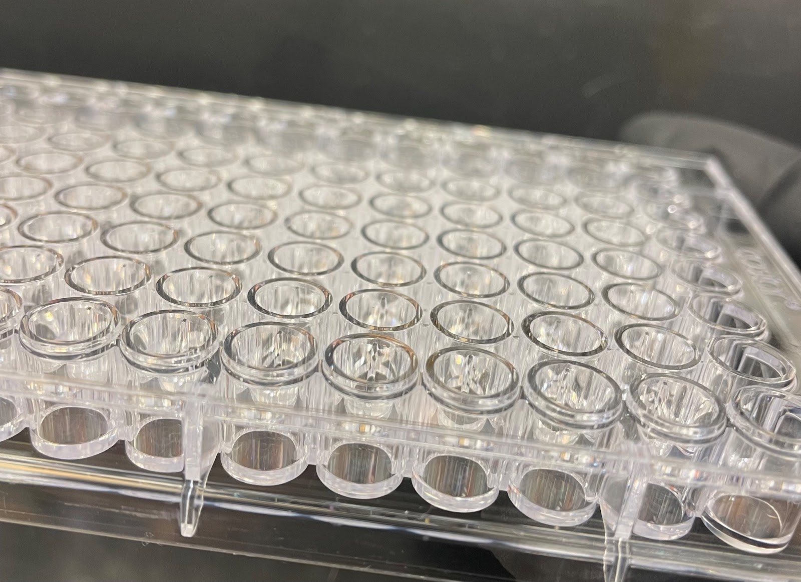
Microtiter plate wells act as small “test tubes” for our microbes to grow in and form biofilms on. An example of what this can look like with a type of microbe called bacteria is pictured below. The bacteria cause the liquid to turn an opaque, yellow color that you can see in some of the wells. After enough time has passed to allow biofilms to form, it’s important to wash off the microbes that are not attached to the plastic plate because those microbes are not a part of the biofilm.

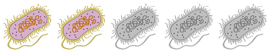
Next, scientists stain the biofilms with a purple dye called crystal violet. After staining, the extra crystal violet needs to be washed off. Then, the plate could look similar to the plate pictured below. We can see in this picture that different wells contain different amounts of crystal violet attached to the biofilms. This tells us that the microbes put into those wells formed better biofilms than the microbes put into the wells with less purple.

The last step of the assay is to add a strong acid or alcohol to the wells to dissolve the crystal violet and then measure the intensity of the purple color, also known as the optical density, with a plate reader. While we can sometimes tell which microbes are better at building biofilms than others by just looking with our eyes, this last step allows us to quantify how dark the purple is in each well. A darker purple color means that more crystal violet stuck to the biofilm, which means that there was more biofilm formed by the microbes.
One reason why microbiologists use crystal violet biofilm assays is as a tool to find proteins important for biofilm formation. Microbiologists can study microbes’ proteins using genetics, the study of DNA and genes. Scientists can delete or change a gene, which is a piece of DNA that contains information needed to create a specific protein, from the microbe’s genome. Then, scientists can see how that gene and its corresponding protein change the way the microbe looks or acts. This is useful in the context of biofilms because if microbiologists can discover which proteins are important for biofilm formation, scientists could develop antimicrobial treatments that target the activity of those proteins. This could prevent the microbes from forming biofilms and make it easier to kill disease-causing microbes.
