by Yasemin Cole
Technique Name: Histology
Fun Rating: 4/5

Difficulty Rating: 4/5
What is the general purpose?
Scientists use histology to study structures within the cell (around 0.01 mm, about 1/10th of the size of a grain of salt). With the naked eye, we can only see 0.1 mm, about the size of a human egg cell. We supplement our vision with microscopes to visualize structures at the microscale, such as at the subcellular level.
Why do we use it?
Histology is the study of small structures of tissues, for example, cells and organelles within a cell. Scientists like to visualize structural changes after changing one variable within a cell. For example, researchers may be interested in visualizing the location of proteins or abnormal shapes of mitochondria within a cell.
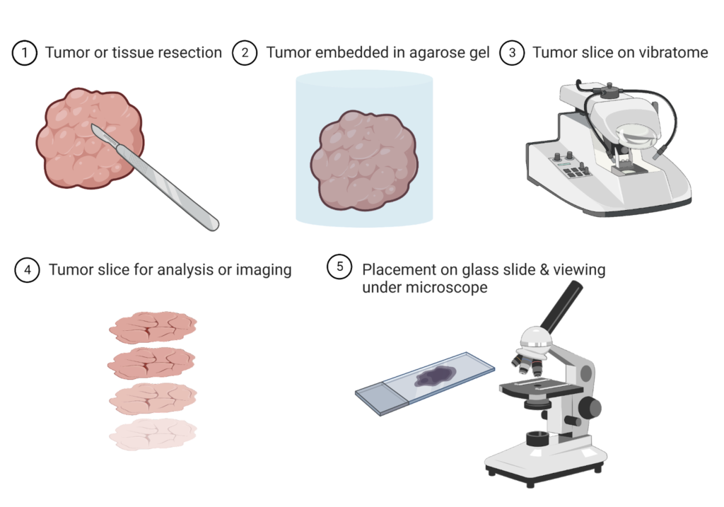
Image created by author using BioRender
How does it work?
- Obtain cells/tissue
Scientists must first collect cells from Petri dishes or tissue samples from a model organism (such as mice, fruit flies, or rabbits) to visualize cells. Depending on the disease or process a scientist is studying, they may use specific types of cells or animal tissues. For example, scientists studying the spread of breast cancer cells may collect tissue from different organs like the liver and lungs to see if the cancerous cells have moved to that region. It is impossible to look at the whole mice under the microscope, so they need to isolate the area of interest.
- Stabilize cells/tissue
Once cells or tissues are isolated, they typically will not be preserved and begin undergoing apoptosis as soon as they are removed from a nutrient source. To prevent the cells from dying, scientists will add a solution that “freezes” the cells or embeds tissue in paraffin wax. Because the tissue is very large, it must be sectioned into small slices and placed onto a glass slide. A review of this process can be found here.
- Visualize cells/tissue
Afterwards, cells can be viewed under a microscope, and scientists can note any changes in the cells or may plan experiments with the cells to explore the protein, DNA, or RNA within the cell. For example, a scientist may want to look at the protein with immunohistochemistry or immunofluorescence, perform fluorescent in situ hybridization to look at the genetics of the cell, or extract RNA.
Histology provides an avenue for exploring a cell’s microstructures and allows scientists to plan future experiments to answer specific scientific questions. It is a fundamental technique in any molecular/cellular biology laboratory using a microscope, reagents, and cell culture or animal tissues.
