by Asjah Wallace
Fun Rating: 4/5

Difficulty Rating: 1/5

What is the general purpose?
The scratch wound assay is a cell culture technique to analyze cell motility (independent, self-driven movement of an organism) over time. It provides insight into cell behavior related to the spread of cancer, cell interactions, and the healing of damaged tissue.
Why do we use it?
This method is easy, cost-effective, and can accommodate a wide range of experimental conditions, such as genetic changes in cells or treatment with therapeutic drugs. Motility is measured based on the ability of cells to migrate into a cell-free gap or “wound” area within a certain period of time. In the broader context of cancer or tissue repair, the time it takes to fill the wound area can indicate the likelihood that cells will mobilize to either invade distant organs or repair tissue.
How does it work?
Cell Preparation
Place your cells in a sterile, multi-well plate and wait until they grow to cover the entire well.
Scratch Formation
There are multiple methods of generating the scratch wound area; however, each technique delivers varying degrees of reproducibility. Manual scratching with the use of a sterile pipette tip can be effective; however, this method creates inconsistent wound areas between samples.
Another technique utilizes a specialized device called a “Wound Maker” that can generate identical wounds in seconds. While this device generates uniform wound areas, the metal pins that create the scratch can become miscalibrated, leading to high replacement costs and inconsistent results.
A third method involves Ibidi well-inserts. Similar to the uniform wound areas generated with the specialized device, Ibidi well-inserts provide an efficient and reproducible method of generating a defined wound area at lower costs. Before plating cells, place the silicon inserts into the desired wells of the cell culture plate. Then, place culture media containing the cells of interest on both sides of the silicon insert. Once cells have grown and covered each side of the well-insert, sterile tweezers are used to gently remove the well-insert, leaving a defined cell-free gap.
Imaging and Analysis
After generating the scratch wounds, images are taken with a brightfield microscope or live-cell imaging system to assess cell motility and wound closure over time (Figures 1-4). Using image analysis software like ImageJ, quantify the wound area at each time point. Then, calculate the percentage of wound closure as (initial wound area-area at defined time point)/(initial wound area).
Representative Images of Scratch Wound Assay
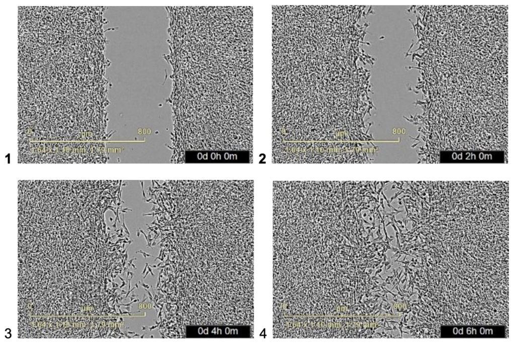
Images taken using Incucyte S3 Live Cell Imaging System.
Figure 1. Initial scratch wound after removal of the Ibidi well-insert.
Figure 2. Image taken at 2-hour timepoint and observe minimal wound closure.
Figure 3. Image taken at 4-hour timepoint and observe mild decrease in wound area
Figure 4. Image taken at 6-hour timepoint and observe significant decrease in wound area
