by Siyao Wang
Fun Rating: 2/5
Difficulty Rating: 4/5
What is the general purpose?
Cryosectioning is a lab method in which tissue samples are rapidly frozen and cut into very thin slices, called sections, for study under a microscope. Freezing helps keep the cells and tissues in their natural shape, making it easier to see their structure and any signs of disease.
Why do we use it?
Researchers often study how different parts of a cell look (morphology) and how they are arranged within the cell. For example, they may want to know how certain neurons are distributed in the brain, or what gut cells look like in the small intestine. By preserving tissue morphology and enabling rapid sample processing, cryosectioning enables high-resolution imaging when combined with immunohistochemistry (IHC).
Outside of a university lab, this procedure is most commonly used in disease diagnosis. After a tissue sample is taken from a patient, a pathologist evaluates the specimen. The pathologist then reports whether the patient’s specimen is diseased or not to aid the determination of future treatments.
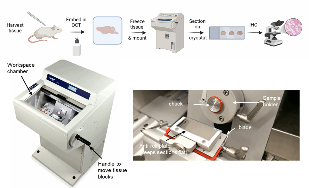
Figure 1: The cryosectioning process. Generated by BioRender by the author.
How does it work?
Cryosectioning usually involves a couple of key steps: preparing the cryostat equipment, preparing frozen samples, and sectioning the samples on the cryostat. OCT (Optimal Cutting Temperature); IHC (Immunohistochemistry)
- Preparing the cryostat: before sectioning samples, the cryostat workspace needs to be cleaned, and a new blade installed to ensure smooth sectioning or slicing of the sample. The chamber needs to be kept at or below -20℃ to avoid the frozen sample from melting.
- Preparing samples: researchers place their tissue samples in a mold (rectangular, square, or circular) and embed the samples in a special media called Optimal Cutting Temperature compound, or OCT for short. OCT is one of the most commonly used media because it holds tissue in place, allows rapid freezing, and provides the support needed to cut thin, intact sections. Once the samples are properly embedded, they will be frozen in a freezer to form a solid block, which is then mounted (or placed) onto a sample holder and sectioned (or sliced).
- Mounting & sectioning samples: the frozen samples are “glued” onto sample holders called chucks (Figure 1) using extra OCT in the cryostat chamber. Because OCT rapidly freezes under low temperature, it’ll firmly attach the sample block to the stub, acting like glue. The chuck will then be inserted into the sample holder, and researchers will section the block by moving the sample toward the blade in an up-and-down motion using the handle. It takes practice and patience for researchers to master this technique to obtain perfect sections.
If you’re interested in learning more about how cryosectioning is done, check out this video: Cryostat Tutorial
