by Yasemin Cole
Fun Rating: 4/5
Difficulty Rating: 2/5

What is the general purpose?
This imaging technique utilizes x-rays to help researchers visualize an organism’s internal anatomy in 3D, allowing them to identify regions of interest for further research. For example, a scientist may be studying the vasculature and wing webbing of a cicada or bird wing and how they have evolved due to environmental selection.
Why do we use it?
We use this imaging technique to assess very small structures (smaller than the size of cells!) inside biological specimens without manipulating them. It can be useful to see the inside of an object without opening or cutting into it. For example, if a scientist is studying cancer in a mouse, they may want to image the whole body to help direct their studies to specific organs of interest, like the liver or brain. Similar to a patient, they may not know where the cancer has spread to, so imaging will help them to focus on an area of interest.
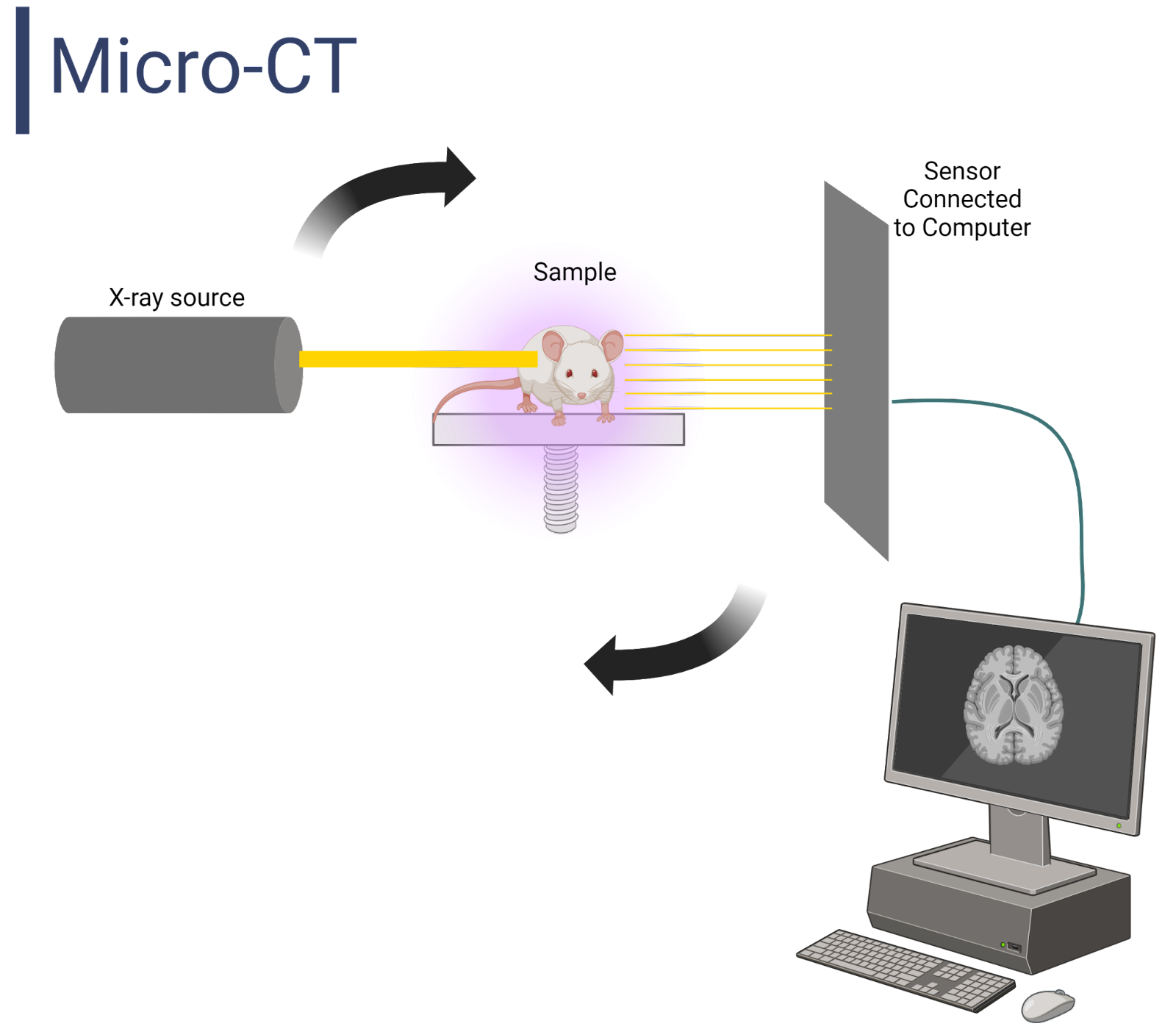
Image created by author using BioRender.
How does it work?
Micro-CT imaging utilizes X-rays to generate images, with slight differences. First, how does a typical X-ray machine in the hospital work? The basis of X-ray images is that dense structures tend to absorb more beams of energy than less dense structures. For example, bone would absorb more beams of energy than muscle. Because of the density differences, X-ray beams will find it difficult to go through a dense object and vice versa. Images are then generated in a black and white gradient, with white representing the densest object. In contrast to typical X-ray machines, computed tomography (CT) uses x-rays that rotate around an object, instead of shooting beams of energy from the front of an object and detecting it at the back. What makes micro-CT different from CT is the ability to visualize structures at the micrometer scale. Before setting up any type of CT scan, care must be taken as X-rays can cause damage with excess exposure due to ionizing radiation.
Sample Preparation:
Micro-CT imaging is typically performed on small biological samples rather than on people and with high quality. Typically, there is very little that scientists need to do to prepare their samples for a micro-CT scan. If a scientist is working on a live mouse, it may be given anesthesia so it does not move in the scanner.
Because we may be interested in looking at two structures with similar densities, we sometimes want to add a contrast agent to the sample before data collection to enhance the density differences in a micro-CT scan. These agents can add “density” to the structure and stop the X-rays from passing through. For example, iodine can be injected into the bloodstream to visualize hollow organs such as blood vessels. Iodine fills the empty space and limits the ability to pass through the tissue, causing a white appearance in the image.
Data Collection & Processing:
In a micro-CT scan, we begin by pointing a narrow beam of X-rays at the sample and rotate the beam 360 degrees. The x-rays that pass through are picked up by a detector 180 degrees behind the sample, then digitized and processed by an attached computer. The generated cross-sectional slices are known as tomographic images, hence the term “computed tomography”. As images are collected, they are digitally stacked on top of each other (called reconstruction) to form a 3D image. What makes micro-CT unique is that each slice generated can be as small as 100 nanometers thick, around one-thousandth the width of a strand of hair.
This process has the advantage of maintaining the sample for future testing. A scientist can complete another scan or can then study an abnormal structure identified by the 3D scan within the sample. You can even 3D print your images! If you want to learn more about this technology you can find more information on the Micro Photonics website.
