by Sarah Angle
Timely and accurate disease detection often determines how effective treatment will be. However, many diseases are difficult to diagnose quickly and without the use of invasive procedures. Traditional diagnostic tools require tissue biopsies, extended laboratory processing, and markers that may be inaccurate. These limitations can delay treatment, reduce patient comfort, and compromise outcomes.
Fluorescent probes offer a promising alternative by enabling rapid, non-invasive, and precise detection of disease biomarkers, or biological “clues” that help doctors understand what’s happening in the body. These probes show visual signals of disease to help doctors decide on treatment.
Figure 1. Activation of a model fluorescent probe after binding to the biological target. Image created by author via BioRender.com.
Fluorescent probes typically consist of three key components: (1) a recognition unit, (2) a fluorophore, and (3) a linker. The recognition unit binds to the biological target, providing the probe with selectivity. The fluorophore serves as the signal-generating element, usually remaining quenched or “off” until activated by the target. The linker connects the recognition unit to the fluorophore and can also influence important properties such as water solubility, circulation time, and overall stability.
The fluorophore can be “turned on” through a variety of reactions, depending on how the probe is designed. Some are activated enzymatically, meaning a disease-related enzyme removes a protective group, which causes the signal to switch on. Others respond to changes in the local environment, such as oxidative stress, shifts in pH, or the presence of specific metal ions. Light-activated probes can even be controlled with precision in space and time.
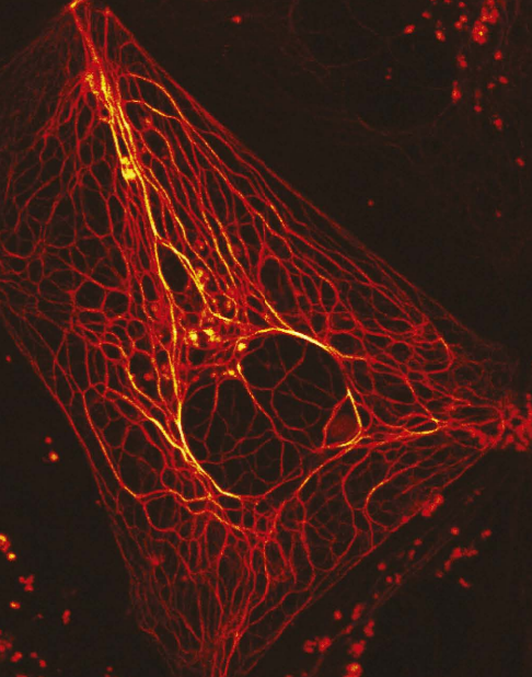
Figure 2. Fluorescent probes are incredibly precise! This one lights up only keratin, the tough protein that gives strength to your hair, skin, and nails.
(Grotjohann T. 2012. Wikimedia Commons. File:RsEGFP2-enables-fast-RESOLFT-nanoscopy-of-living-cells-elife00248f002.jpg – Wikimedia Commons)
Fluorescent probes have demonstrated promise across a wide range of medical applications. In oncology, they are used to highlight tumors during surgery, helping surgeons distinguish cancerous and healthy tissue in real time. In infectious disease, probes can rapidly detect pathogens, or harmful germs, by binding to unique proteins, offering faster results than traditional culture-based methods that require growing the pathogens outside of the body. Probes are also being developed to monitor metabolic changes, track drug delivery, and visualize immune system function.
