by Liseth O. Barrionuevo
CANCER CELL CULTURE
What is the general purpose?
The general goal of cancer cell culture is to study cancer biology. Specifically, by utilizing cancer cell lines, scientists can investigate different aspects of cancer cells from various cancer types.
Why do we use it?
Each scientist is going to use this technique according to the specific goals of their project, so there are plenty of lab tests that can be done through cancer cell culture; for example, a scientist could observe how cancer cells grow, interact, and/or react to treatment in vitro (in a plate under controlled conditions). In cancer research, this technique is one of the first steps for testing and validating hypotheses because it is generally more affordable than in vivo methods involving living organisms such as mice. In this sense, cancer cell culture is essential to generate data that could potentially lead to subsequent animal experiments – for instance, cancer cell injections in lab mice.
So, how does this all relate to soup? The same way your soup provides you with a variety of nutrients such as proteins, carbs, vitamins, and much more, cells also need a rich “broth” with the right nutrients, which scientists call culture media, for their specific cell type to thrive in a controlled environment in terms of temperature, CO2, and oxygen levels.


Figure 1: Popular winter soups. Sources: https://unsplash.com/photos/a-bowl-of-soup-VUr9FsuIgqc; https://unsplash.com/photos/soup-in-bowl-fxJTl_gDh28
To explain more about how the process works, let me walk you through a story with you as the protagonist. First, imagine yourself in your dream lab. Put on your lab coat, gloves, a bouffant cap, and a disposable mask. We are situated and all dressed up for our adventure, let’s roll! Let’s gather our materials and place them in the laminar flow hood (a cabinet-shaped environment) to ensure as much sterility as possible (Figure 2). The flow hood is essential for cell culture, as air passes through integrated filters to maintain a constant airflow that prevents outside contamination from entering the workspace.

Figure 2: Laminar flow hood for cell culture. Source: https://commons.wikimedia.org/wiki/File:Laminar_Flow_Cabinet_for_Tissue_Culture.jpg
The process of cell culture could be complex, depending on the goal of your experiment. However, I will walk you through the most general workflow. First, to start, we need to “wake up” our cancer cell lines, which are established cancer cells of a specific type that can be purchased for your experiments. These cancer cell lines come to your lab frozen in storage vials (like the small plastic tubes with orange caps in Figure 3), so what I meant by “waking them up” is that we are going to take them out of the freezer on a bed of ice, add some culture media into each vial and transfer the whole content to a culture dish or bottle, which already has fresh culture media ( the rich “broth” we talked about before).
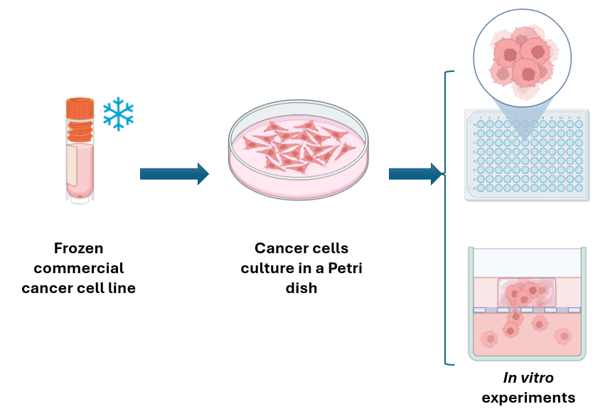
Figure 3: General workflow of cell culture. The author´s original image created in Biorender.
Now that you have your cancer cells in a culture dish or bottle, you need to put them in an incubator, which provides a controlled environment generally set to 37 Celsius, 5% CO2, and >95% humidity. Make sure to check your cells within the next 24 hours and replace the culture medium with a fresh supply, depending on your specific cells’ needs. For the next steps, it depends on what assays (specific experiments) you need to do, but it’s a good idea to prevent your cells from covering all the surface of the culture dish, instead it is recommendable to keep them below 70% to 80% of confluency (which refers to the percentage of a culture dish covered by cells, check this out to know more: https://ncdnadayblog.org/2023/05/12/confluency/) for your subsequent experiments.
Great, we are all set for now. Wow, you have done a lot today, congrats! Great job getting a general sense of cancer cell culture. Stay tuned for more techniques in our next posts.
